What are hemangiomas and vascular malformations?
- Hemangiomas and vascular malformations are noncancerous growth of blood vessels. They show up at birth or soon after birth. They’re also called birthmarks.
- The cause of these growths is often not known. They may run in family.
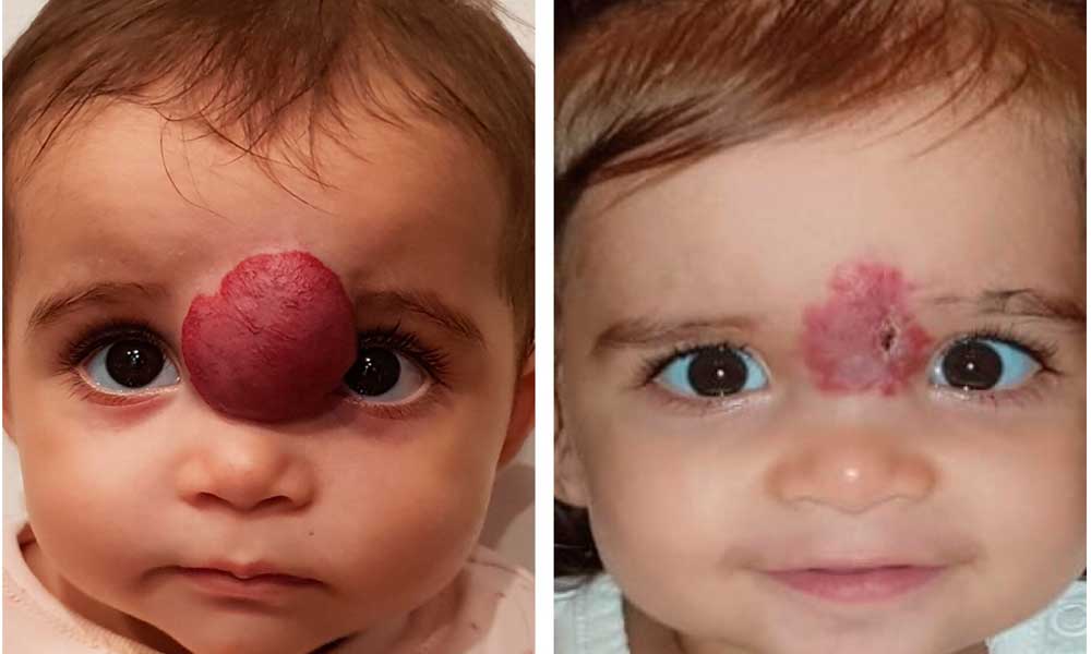
- Hemangiomas often start as faint red marks. Then they grow quickly after birth. Then they start to shrink. They may go away without treatment.
- Vascular malformations grow slowly throughout life. They don’t shrink. They usually require treatment. These lesions sometime infiltrate normal tissue which makes them very difficult to manage
Vascular malformations are of 5 types. They are:
- Port wine stains (superficial red or purple in color)
- Venous malformations ( slow flow lesions)
- Lymphatic malformations ( previously called cystic hygroma” or “lymphangioma”).
- Arteriovenous malformations (AVM) – high flow lesion.
- Mixed malformations, a combination of any of the other types
How are hemangiomas and vascular malformations diagnosed?
The diagnosis will be based on physical appearance and how it changes over time.

Most hemangiomas are in the head or neck area, but they can occur anywhere in the skin, mucous membranes, or internal organs. Most will keep growing for the first 3 to 5 months of life. Then they start to shrink. Almost 50% disappear by the age of 5 yrs and the vast majority are gone by age 10. Up to 50% of children with hemangiomas may have skin issues such as scarring, skin discoloration, and tissue wasting (atrophy).
Venous malformation may present as a deep mass. The protrusion may be the only presenting symptom. The overlying skin may appear normal or possess a bluish discoloration. With more cutaneous involvement, the lesions appear darker blue or purple
They are compressible and swell when the region is dependent or there is an increase in hydrostatic pressure such as during a Valsalva maneuver. With time, pain and swelling may occur with the formation of phleboliths (calcified thrombi), or small clots, secondary to trauma or venous stasis.
Lymphatic malformations Local infections close to the course of lymphatic drainage will cause LM to swell, protrude, and sometimes become painful. This is a hallmark of an LM versus other vascular anomalies that do not present in this fashion.
Arteriovenous malformations (AVMs)– Puberty and trauma trigger the growth of the lesion They are infiltrative causing destruction of local tissue and often life-threatening secondary to massive bleeding. Palpable warmth, pulse, or thrill due to its high vascular flow is the hallmark. The overlying skin may have a well-demarcated blush
What are the complications of hemangiomas and vascular malformations?
These conditions can be life-threatening if they’re large or affect a child’s airway
A hemangioma and AVM can also be serious if it has uncontrollable bleeding.
Depending on where growth is located, it may cause physical problems like may have trouble moving part of his or her body.
When involving the skeletal framework in this area, LMs often cause osseous hypertrophy leading to dental or extremity abnormalities.
Diagnosis –
Doppler Ultrasound Study is the basic and cost-effective study to show the type of vascular channels whether lymphatic, venous or arterial and also the extent of the lesion.
MRI /MRA / CTA– In the case of internal lymphatic or arterial malformation, these modalities are used to confirm the diagnosis, identify cystic or vascular architecture, and determine the extent of disease and its adjacent infiltration.
Arteriography can show the central nidus necessary for intravascular treatment.
How are hemangiomas and vascular malformations treated?
Small hemangiomas often shrink on their own. These usually don’t need treatment.
If the hemangioma is near the eyes or airway, it may need to be treated. Almost all vascular malformations and nearly 40% of hemangiomas eventually require intervention.
Treatment Options –
For Small and superficial lesions —
- Intralesional Steroid medicines
- An oral medication beta blocker has been safely used to treat symptomatic hemangiomas.
- Superficial Laser therapy. This is used for port wine stains.
- Residual erythema and telangiectasias that frequently remain in involuted hemangiomas are best treated by selective photothermolysis
For large lesions —
- Embolization of the blood vessels of hemangioma / arteriovenous malformation – This is the most effective and patient-friendly treatment option for large or symptomatic lesions. Is done on Daycare basis under local anesthesia and depending upon the size of the lesion, one or more sittings may be required.
- Sclerotherapy – Injection of clotting (sclerosing) medicine like ethanol, bleomycin, OK-432, and doxycycline for venous and lymphatic malformations. These lesions infiltrate normal tissue which makes them very difficult to manage
- Surgical Removal – Used for very large lesions but failure to completely excise LM and VM often leads to recurrence. Preoperative sclerosant/embolization can be used prior to excision (24–48 hours) to decrease surgical risk.
Non-surgical treatment – Embolization of the blood vessels of hemangioma / arteriovenous malformation:
This is the most effective and patient-friendly treatment option for large or symptomatic lesions. Is done on Daycare basis under local anesthesia and depending upon the size of the lesion, one or more settings may be required.
This Non-surgical catheter directed embolization is an outpatient treatment. The specifically trained interventional radiologist makes a tiny nick in the skin at the groin or neck using local anesthesia and a thin catheter is passed and directed to the main artery supplying the AVM. With contrast dye injection, interventional radiologist maps out exactly the extent and caliber of vessels.
By using specialized glue, PVA particles and coils of appropriate size, embolization of the nidus is done (artery supplying the AVM). The material used for embolization depends on the features of AVM. The whole non-surgical treatment procedure is painless and takes about 30 minutes. The tube is removed and no stitches are needed.
Patients are observed for a few hours and go home the same day. Patients often return to work the next day.
Dr. Pradeep Muley is trained in the USA and Singapore and has performed over 20,000 non-surgical treatments for various diseases like uterine fibroids, uterine adenomyosis, un-operable liver tumor, liver abscesses, varicose vein, brain aneurysm and vomiting of blood from lungs and stomach. He runs the VARICOSE VEIN AND FIBROID CLINIC AT FORTIS HOSPITAL, VASANT KUNJ, NEW DELHI & created INDIA’S 1ST UTERINE FIBROID CLUB. He has treated the maximum number of fibroid patients in India through the innovative Uterine Artery Embolization method.
For more in-depth information on various non-surgical treatments available-
Mail Us: muleypradeep@hotmail.com or Visit to Website: – www.indianinterventionalradiology.com
Phone or Whatsapp: +91-9810492778